
Tiny fragments of plastic appear to be found wherever we look.
Humans have been making ever-increasing quantities of this material since the 1950s, so it is hardly surprising that these persistent polymers are widespread throughout the environment.
As plastic degrades into progressively smaller pieces, it can also enter our bodies.
Concern about microplastics may already be widespread, yet we still understand very little about their effects on health after they enter the human body.
A new study, led by scientists at Vrije Universiteit Amsterdam and published in Molecular and Cellular Endocrinology, shows that some micro- and nanoplastics may cross the placental barrier and alter steroid hormone production during pregnancy.
Scientific research has already identified micro- and nanoplastics in human blood, placentas, amniotic fluid and meconium. One investigation of 30 human placentas detected microplastics in every sample.
Finding these particles near a developing fetus is worrying. During these early stages, when the framework of a new human body is being established, disruption of any kind could have serious effects. However, it remains unclear whether plastic particles genuinely interfere with fetal development in humans.
Micro- and nanoplastics in pregnancy
Research in mice found that minute plastic fragments inhaled by a mother during pregnancy could be present in her offspring’s organs for up to two weeks after birth, although the study did not exclude the possibility that the offspring obtained the particles through other routes.
A separate mouse study indicated that exposure to micro- and nanoplastics in pregnancy has the "potential to disrupt fetal brain development, which in turn may cause suboptimal neurodevelopmental outcomes".
The latest work, headed by environmental health and toxicology researcher Jeske van Boxel, brings researchers closer to understanding the possible effects of these plastics in human pregnancies.
As van Boxel and colleagues note, major anatomical differences between mice and humans, particularly during pregnancy, "indicate that micro- and nanoplastic placental translocation and effects on steroidogenesis during pregnancy in rodents may not be directly translatable to humans".
The authors also write that "existing human placental in vitro models do not fully capture the complex maternal-fetal interactions in steroidogenesis and structural arrangement,".
A human placental barrier model
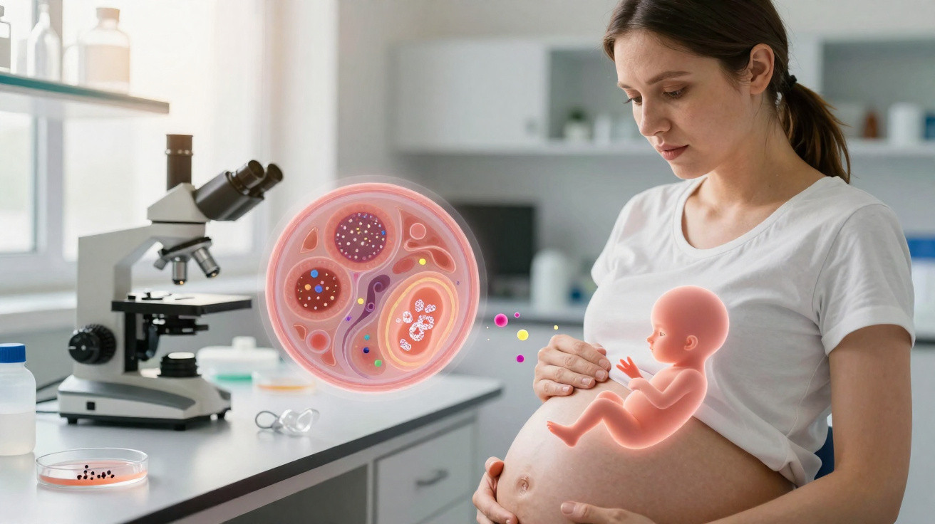
To address this, the researchers created a ‘petri dish’ model containing three layers of distinct living human cells. The model recreates the structure of the barrier layers in a human placenta.
Its first layer comprises BeWo b30 cells, which share characteristics with the epithelial tissue covering placental villi. The second consists of HUVEC cells isolated from a human umbilical-cord vein, while the third contains H295R cells originating from a human adrenal gland.
The BeWo b30/HUVEC layers were selected to represent the placenta’s physical barrier. The H295R layer represents the main hormone-producing organ within the placenta, called the fetal adrenal.
After establishing the system, the team introduced several types of micro- and nanoplastics to its ‘outer layer’. They then examined which particles travelled through the cells and how those particles influenced the hormones produced by the cells.
Over 72 hours, most polymers passed through every membrane layer, although some did so more readily than others. Polyamide was the only type not detected in measurable quantities on the far side of the membrane.
Fluorescence microscopy showed that more plastic particles were embedded across the first two cell layers, but not in the H295R cells.
Steroid hormones and plastic exposure
Even before plastics were introduced, the team detected 19 different steroid hormones in the three-cell model. Although absolute hormone levels differed somewhat from those in pregnant humans in vivo, comparable key steroidogenic pathways were active in the study’s tri-culture model.
The researchers could then assess the changes that followed the addition of plastics.
Exposure to polymers caused a shift in the cells representing the outer part of the model placenta. Several plastic types reduced etiocholanolone, while exposure to polystyrene affected oestrogen levels.
Given the importance of steroid hormones during pregnancy, the researchers say more investigation is required to determine the mechanisms behind these findings.
"Our study shows that several types of micro- and nanoplastics can cross a human placental model and influence hormone production," van Boxel says.
"While this does not directly mean they cause harm during pregnancy, it highlights the urgent need to better understand how these particles may affect fetal development."
The research was published in Molecular and Cellular Endocrinology.
This article was fact-checked by Rachel Garner and edited by Peter Dockrill. While we take pride in our process, we are only human. If you notice an error, please let us know.



Comments
No comments yet. Be the first to comment!
Leave a Comment